Rapid EV Concentration Measurement Without Staining
Counting extracellular vesicles by fluorescence works, and for questions about identity it is indispensable. But when the question is simply how many particles are in this tube, a labelling step adds cost, hours of handling and — more importantly — a set of biases that are easy to overlook because they are built into the method rather than into the sample.
Rapid EV concentration measurement without staining is therefore not a shortcut. It is a different measurement, with different failure modes, and for concentration specifically it is often the more defensible one. This page explains where labelling distorts a count, why dilution cascades matter as much as the label itself, and what a label-free per-particle count does and does not tell you. For the broader characterisation picture, see our overview of extracellular vesicle characterization.
Where fluorescent labelling biases a concentration measurement
You count what you stained, not what is there
An antibody-based approach counts vesicles carrying the epitope you targeted. Marker expression across a vesicle population is heterogeneous, and a tetraspanin-negative subpopulation is not counted at all. As a measure of total particle concentration, a marker-restricted count is a lower bound whose relationship to the true number depends on biology you usually do not know in advance.
Generic dyes stain more than vesicles
Lipophilic and membrane dyes avoid the marker-selectivity problem but create another: they label lipoproteins and membrane fragments as readily as vesicles, and unbound or self-aggregated dye can itself produce events above threshold. Free-dye background is a well-known artefact source, and it inflates rather than depresses the count.
Labelling requires handling, and handling changes the sample
Incubation, washing and removal of unbound label each cost vesicles — adsorbed to tube walls, lost in a wash, or aggregated by a buffer change. The measurement is performed on the preparation that survived the protocol, not the one you started with.
Weak scatterers stay weak
Fluorescence relaxes the scattering-detection problem but does not remove the size-dependent one: the number of accessible epitopes or the amount of incorporated dye falls with vesicle surface area, so the smallest vesicles remain the hardest to detect. The detection floor moves; it does not disappear.
The problem underneath the label: working concentration range
Every single-particle counting method has a window within which the count is valid. Too concentrated, and more than one particle occupies the detection volume at once — two vesicles are recorded as one larger event, so concentration is under-reported and size over-reported. Too dilute, and too few events accumulate for a statistically meaningful number.
Staying inside that window normally means a dilution series, and this is where a surprising share of real-world error originates. Each dilution step carries its own pipetting uncertainty; the factors multiply; and because vesicles adsorb to surfaces and can dissociate on dilution, a diluted sample is not always a faithfully scaled version of the original. When the required dilution is large, uncertainty in the final answer is dominated by the preparation, not the instrument.
Reducing the number of dilution steps is therefore one of the most effective ways to improve a concentration measurement — independent of any labelling question.
Label-free per-particle counting with interferometric light microscopy
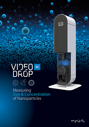
Interferometric light microscopy (ILM), the principle behind the Videodrop SC, detects each nanoparticle individually through the interference between the light it scatters and the incident beam. It returns a number-based size distribution and a concentration in particles/mL from a single 5–10 µL drop, in under a minute, with no dye, no antibody, no incubation and no wash.
What that changes in practice:
- No marker selectivity. Every detected particle above the threshold is counted, whatever it expresses. For total particle concentration, this is the quantity you actually wanted.
- No free-label background, and no conjugation chemistry to validate or re-validate per lot.
- Minimal dilution. The working range of 108–1010 particles/mL accommodates many EV preparations directly, so the dilution cascade largely disappears — and with it its error.
- The sample survives. The measurement is non-denaturing and label-free, so material can be recovered and used downstream.
- Aggregates are visible, not inferred. Live imaging shows debris and aggregates up to 10 µm, so you can see when a count should not be trusted rather than discovering it later in a distorted distribution.
- No settings to tune and no calibration, which removes inter-operator variation in the count itself.
The approach has been used in clinical research on plasma vesicles, including a study of glioblastoma patients (Sabbagh et al., Scientific Reports, 2021) and longitudinal monitoring of vesicles from metastatic breast cancer patients under treatment (Richard et al., Journal of Extracellular Biology, 2024).
What a label-free count does not tell you
Two limits must be stated plainly, because they determine whether this is the right measurement for your question.
It does not establish identity. A light-based counter cannot distinguish a vesicle from a lipoprotein or a protein aggregate of similar size. This is exactly why the consensus guidelines published as MISEV2023 require a particle count to be accompanied by molecular evidence that the particles really are EVs. Label-free counting replaces fluorescence for quantity; it does not replace it for identity, and marker-based methods remain necessary for that.
It has a detection floor of around 80 nm. Since exosomes span roughly 30–150 nm, the smallest vesicles are outside the window. The peer-reviewed comparison by Sausset et al. (2023) documented this directly: a higher detection threshold than particle tracking, offset by greater speed, simpler handling, fewer consumables and less small-particle masking in highly polydisperse samples. In practice this makes the method strong for relative quantification, process monitoring and longitudinal follow-up — where consistency between measurements matters more than absolute completeness — and weaker as a claim of total vesicle number.
A defensible workflow
- Count label-free for concentration, in as few dilution steps as possible.
- Run a buffer blank and subtract background before interpreting any value.
- Measure in replicate and report dispersion, not a single mean.
- Confirm identity separately with orthogonal molecular evidence, per MISEV2023.
- Report the method and its detection threshold alongside the number — a concentration without its method cannot be compared between laboratories.
Key takeaways
- Marker-based counting measures a labelled subpopulation; generic dyes add lipoprotein and free-dye artefacts.
- Much real-world error comes from the dilution cascade, not the detector — fewer steps means a better number.
- Label-free per-particle counting gives total particles/mL in under a minute from a single drop, with no incubation or wash.
- It does not establish EV identity and does not reach below ~80 nm; keep orthogonal molecular evidence in the workflow.
Need particles/mL without a staining protocol? See how the Videodrop SC counts and sizes vesicles label-free in a single drop — request a demonstration.
Choose your application



Choose your product
Measuring
Size & Concentration
of Nanoparticles
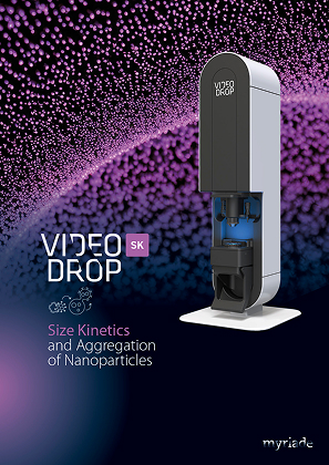
Size Kinetics
and Aggregation
of Nanoparticles