Phage Titer Without Overnight Incubation: What a Physical Count Can and Cannot Replace
The double-agar-overlay plaque assay has been the reference for bacteriophage quantification for the better part of a century, and for good reason: it counts infectious units, which is usually what matters biologically. It also takes overnight. During phage isolation, purification or process development, that latency sets the pace of the entire project — you make a decision, wait a day, then make the next one.
A phage titer without overnight incubation is possible, but only by measuring something different: total physical particles rather than plaque-forming units. Understanding that difference is what makes the fast measurement useful instead of misleading.
Before anything else: does your phage fit?
This determines whether the rest of the page applies to you, so it comes first rather than last.
Optical particle counting by interferometric light microscopy has a detection threshold of around 80 nm. Bacteriophages span a very wide size range, and a substantial number of lytic phages — including many small tailed and small icosahedral phages — have capsids at or below that bound. For those, the method does not apply, and no workaround changes that.
When assessing your own phage, consider the whole particle rather than the capsid diameter alone: a tailed phage with a modest capsid but a long tail presents a larger effective scattering object than its capsid dimension suggests, and detectability depends on refractive index as well as size. The reliable approach is empirical — measure a known-titer preparation of your phage and see whether the count is credible before designing a workflow around it. For very small phages, particle tracking or electron microscopy remain necessary.
Two different numbers
The plaque assay and a physical count are not competing estimates of the same quantity, and treating them as such causes real confusion in the literature.
| Plaque assay (PFU/mL) | Physical particle count (particles/mL) | |
| What is counted | Particles able to infect and lyse the host | All particles above the detection threshold |
| Turnaround | Overnight or longer | Under a minute |
| Host required | Yes — and results depend on the strain used | No |
| Counts defective particles | No | Yes |
| Counts non-phage particles | No | Yes — debris and vesicles are included |
| Aggregates | An aggregate forms one plaque | Visible directly in the image |
| Consumables | Plates, agar, host culture, dilution series | None — label-free, no calibration |
The ratio between the two is itself informative. A particle-to-PFU ratio far above unity means a large fraction of your particles are non-infectious, defective, aggregated — or not phage at all. Tracking that ratio across a purification is often more revealing than either number alone, and it is a measurement you simply cannot make without both.
Where a fast physical count earns its place
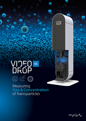
Interferometric light microscopy, the principle behind the Videodrop SC, measures particle concentration and a number-based size distribution from a single 5–10 µL drop in under a minute, label-free and non-denaturing, with live imaging of aggregates and debris up to 10 µm. Its relevance to phage work is not hypothetical: bacteriophages were one of the two sample types in the peer-reviewed comparison by Sausset et al. (2023), which placed the method alongside particle tracking and found it faster, easier to handle, requiring fewer consumables and less prone to small-particle masking in highly polydisperse samples — with a higher detection threshold as the trade-off.
Given that profile, the applications where it genuinely helps are specific:
- Monitoring purification in real time. Concentration steps, precipitation, density gradients and filtration can each be assessed immediately, with a recovery figure per step rather than a single overnight number at the end.
- Detecting aggregation. Phages aggregate on concentration, buffer exchange and storage, and a plaque assay hides it completely: an aggregate of a hundred particles produces one plaque, so aggregation reads as a titer drop indistinguishable from a real loss. Seeing the aggregates directly separates those two explanations.
- Screening lysate conditions. Comparing host strains, media, multiplicities of infection or harvest times becomes a same-day experiment across many conditions instead of an overnight one across a few.
- Stability studies. Repeated readings on the same preparation over time, at 5–10 µL a reading, make storage and freeze-thaw studies cheap.
- Rapid dilution targeting. A fast physical count tells you approximately where a sample sits before you commit an overnight plaque assay to the wrong dilution range — a small use that saves a surprising amount of repeated overnight work.
What it cannot do
Three limits, stated plainly, because a page that oversold this would waste your time and ours.
It does not measure infectivity. Only a plaque assay or an equivalent infection-based method tells you how many particles can kill a host. For therapeutic potency, release testing and any host-range question, the plaque assay remains necessary and this measurement does not substitute for it.
It is not phage-specific. Membrane vesicles, cell debris and protein aggregates in a crude lysate are counted as particles. The number is most meaningful on purified or partially purified material — on crude lysate, treat it as a trend, not an absolute phage titer.
It has a size floor. As set out at the top: below roughly 80 nm the method does not apply, which excludes a meaningful share of phages.
A workable combination
- Verify applicability first on a known-titer preparation of your own phage.
- Use the physical count for speed — process steps, condition screens, aggregation checks, dilution targeting.
- Use the plaque assay for truth — infectious titer, potency, host range.
- Track the particle-to-PFU ratio across the process; it is the most useful number neither method gives alone.
- Report both, with the method alongside each value.
Key takeaways
- A physical count and a plaque assay measure different things; the fast one does not replace the slow one.
- The ~80 nm detection floor excludes many phages — verify with your own preparation before building a workflow.
- Where it applies, it turns purification monitoring, condition screening and stability work from overnight into same-day.
- Its unique contribution is aggregation visibility, which a plaque assay cannot provide at all.
Working on phages above 80 nm? See how the Videodrop SC counts particles and reveals aggregates in a single drop — request a demonstration and test it on your own preparation.
Choose your application



Choose your product
Measuring
Size & Concentration
of Nanoparticles
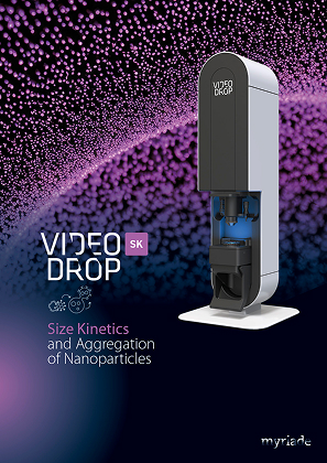
Size Kinetics
and Aggregation
of Nanoparticles